Computer-assisted corneal topography: high resolution graphic presentation and analysis of keratoscopy. Corneal topography of pellucid marginal degeneration. Maguire LJ, Klyce SD, McDonald MB, Kaufmann HE. Topographic analysis in pellucid marginal corneal degeneration and keratoglobus. Su di un caso di degerazione marginale de la cornea-varieta inferiore pellucida-stodie clinico ed istologico. Degenerescence marginale pelluside se la cornee. Arch Ophthalmol 1978 96: 1217–1221.įrancois J, Hassens M, Stockmans L. Topographical analysis is an invaluable tool that may help confirm a clinical suspicion. They need to be recognised and differentiated from each other.

Peripheral corneal thinning disorders are a group of distinct entities with differing implications for management. To the best of our knowledge, this is the sixth such patient reported so far.
#PELLUCID MARGINAL DEGENERATION BELLINGHAM SERIES#
Another series by Biswas et al 9 describes 16 patients with pellucid marginal degeneration, of which it was unilateral in three patients. The first was in an elderly black patient, while the second was an Indian patient similar to the case described. Two cases of unilateral isolated PMD have been reported so far by Wagenhorst 7 and Basak et al 8. Though bilateral conditions do present asymmetrically in degree or time period, the left eye in this case has not shown any sign of the disease 11 years after the right eye started becoming symptomatic. The case reported highlights the rare occurrence of unilateral PMD. It also needs to be differentiated from peripheral corneal disorders associated with inflammation such as Terrien's marginal degeneration, Mooren's ulceration, and ulcers associated with connective tissue disorders.

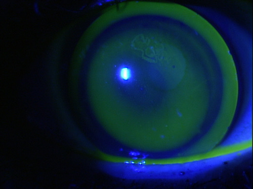
These characteristic features help to differentiate this condition from other noninflammatory corneal thinning disorders such as keratoconus, posterior keratoconus, and keratoglobus. 4, 5 The topographic pattern described is distinctly different from that seen in keratoconus, in which a small area of high corneal power is surrounded by concentric bands of low corneal power. 4 This results in a relatively steep contour approximately 90° away. Stromal thinning is known to cause corneal flattening over the area of tissue loss, and steepening at the border of unaffected tissue. Topographic analysis in our case revealed a characteristic bow-tie appearance of marked against-the-rule astigmatism oblique-inferiorly, without peripheral steepening. This rare entity has been postulated to be an abnormality of the connective tissue, 2, 3 but the exact pathogenesis is still unknown. Pellucid marginal degeneration is a bilateral, slowly progressive condition classically described between the second and fourth decades. The left eye was normal, with no evidence of corneal thinning, striae, or abnormal protrusion ( Figure 1, bottom). The intraocular pressure by applanation tonometry was normal in both eyes. The rest of the anterior and posterior segment was normal. There was no evidence of iron lines, lipid deposition, or vascularisation. The portion between the thinned area and limbus was normal in thickness. On slit-lamp biomicroscopy, the cornea in the right eye showed an irregular contour with a thin band inferiorly, approximately 1.5 mm in width and 2 mm from the limbus, with bulging above the thinned zone ( Figure 1, top). Ophthalmic examination revealed best-corrected visual acuities of 6/18 OD with −1.0/−13.50 D×110° and 6/6 OS unaided. There was no history of redness, pain, or use of ophthalmic medication during this time. Case reportĪ 46-year-old healthy Indian man presented with painless progressive diminution of vision in the right eye for 6 years. Described here is such a case, in which the other eye was normal. Unilateral isolated PMD is extremely unusual, and corneal topography provides valuable clues in suspected cases. 1 It is differentiated from other peripheral corneal thinning disorders such as Terrien's marginal degeneration and Mooren's ulcer by the absence of vascularisation, lipid infiltration, or corneal ulceration.

Pellucid marginal degeneration (PMD) is a rare bilateral corneal disorder characterised by thinning in the peripheral portion of the inferior cornea with marked steepening just superior to the thinned zone.


 0 kommentar(er)
